Spermatic Cord Histology

And because this is fetal tissue the processus vaginalis an extension of the peritoneal space.
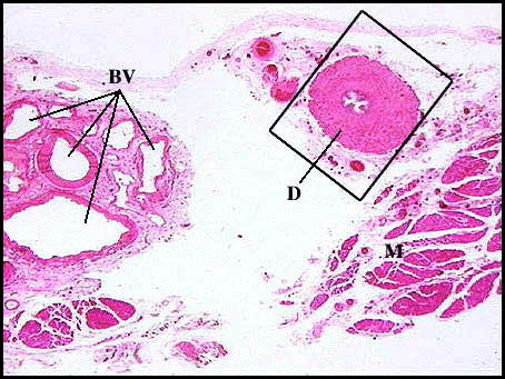
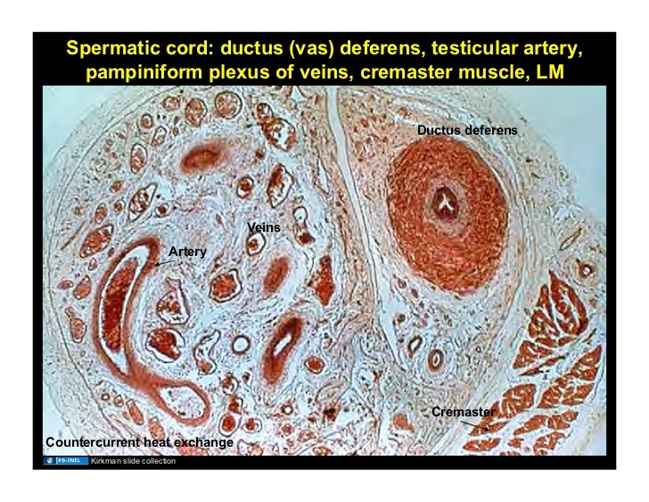
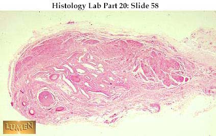
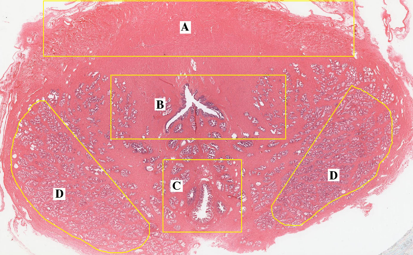
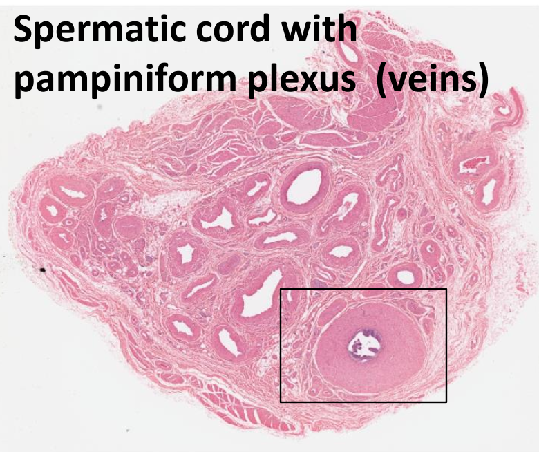
Spermatic cord histology. Its serosal covering the tunica vaginalis is an extension of the peritoneum that passes through the transversalis fascia. The pampiniform plexus of veins. 10x 1 of 5. Ductus deferens continues as the ejaculatory duct in the prostate gland.
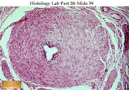
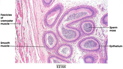
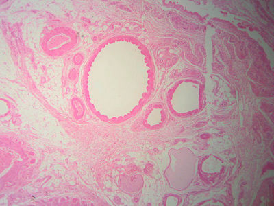
The ductus deferens with three layers of smooth muscle. Spermatic cord varicocele general. Surrounding connective tissue and nerves. The spermatic cord is the cord like structure in males formed by the vas deferens ductus deferens and surrounding tissue that runs from the deep inguinal ring down to each testicle.
Each cord is sheathed in connective tissue and contains a network of arteries veins nerves and the first section of the ductus deferens through which sperm pass in the process of ejaculation. Thought to be due to increased testicular temperature. This section of dentaljuce has over 400 histological slides showing tissues from all organ systems in their healthy state. Each testicle develops in the lower thoracic and upper lumbar region and migrates into the scrotum during its descent it carries along with it vas deferens its vessels nerves etc.
The spermatic cord traversing the inguinal canal is composed of the ductus deferens and its surrounding connective tissue nerves and blood vessels including the testicular artery and the pampiniform plexus of veins. Large varicoceles associated with infertility. The testicular artery with its thick muscular wall.


