Spermatic Cord Anatomy Ct

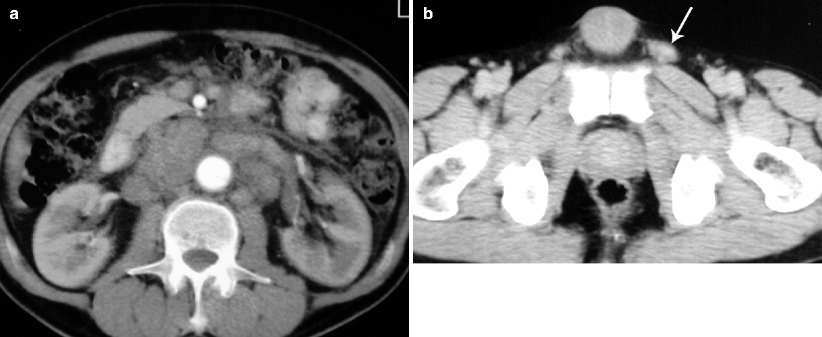
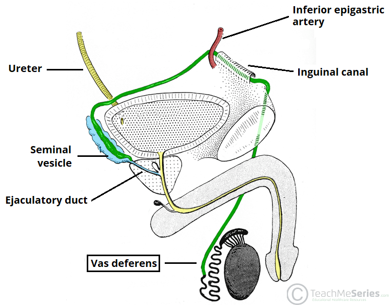
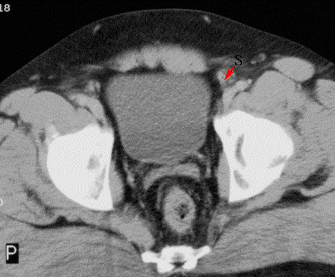
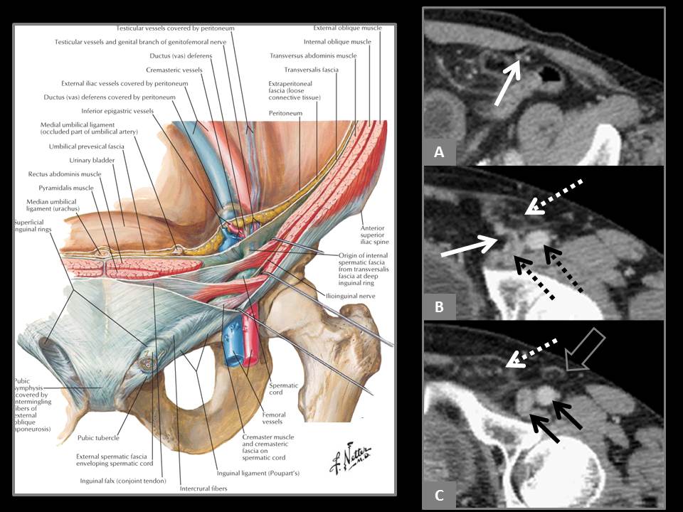
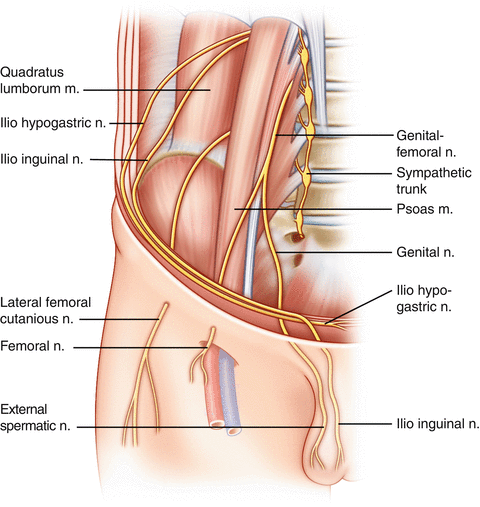
In males the ic transmits the spermatic cord which includes the vas deferens the testicular artery and the genital branch of the genitofemoral nerve from the pelvic cavity to the scrotum fig 4.
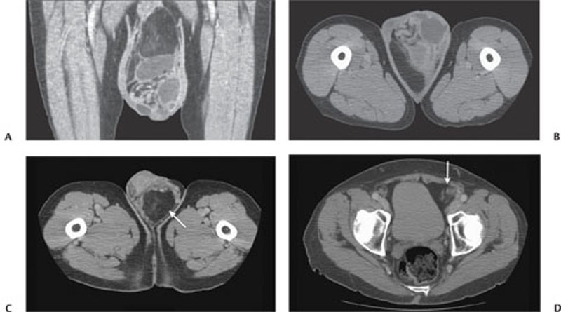
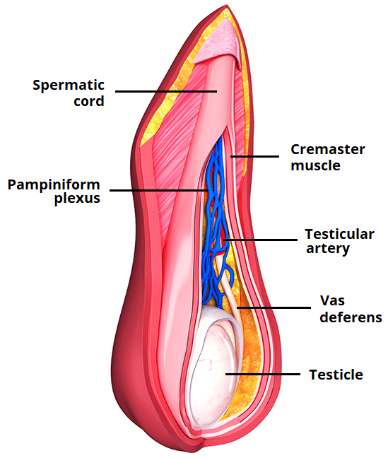
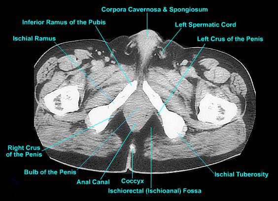
Spermatic cord anatomy ct. Benign processes spermatic cord lipoma lipomas of the spermatic cord are usually inci. The spermatic cord is composed of arteries veins lymphatics nerves and the excretory duct of the testis. Its serosal covering the tunica vaginalis is an extension of the peritoneum that passes through the transversalis fascia. Start studying inguinal and spermatic cord anatomy.
In females the ic transmits the round ligament of the uterus and the ilioinguinal nerve to the labia majora 1. Most present as painless slow growing masses and can be mistaken for inguinal hernias. This opening is located laterally to the inferior epigastric vessels. Gross anatomy course the spermatic cord arises at the deep inguinal ring passes through the inguinal canal and exits.
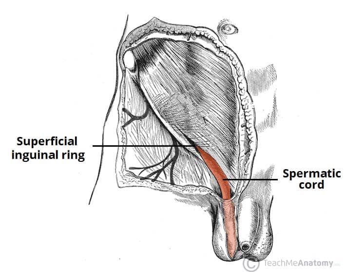
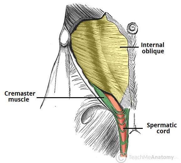
The spermatic cord is the cord like structure in males formed by the vas deferens ductus deferens and surrounding tissue that runs from the deep inguinal ring down to each testicle. Spermatic cord liposarcomas are the most common malignant tumor of the spermatic cord. Treatment is orchiopexy 8. The spermatic cord is the tubular structure that suspends the testes and epididymis in the scrotum from the abdominal cavity.
They are usually well differentiated and spread by local extension. The cord passes through the inguinal canal entering the scrotum via the superficial inguinal ring. It continues into the scrotum ending at the posterior border of the testes. Spermatic cord the scrotum or scrotal sac is a part of the external male genitalia located behind and underneath the penis.
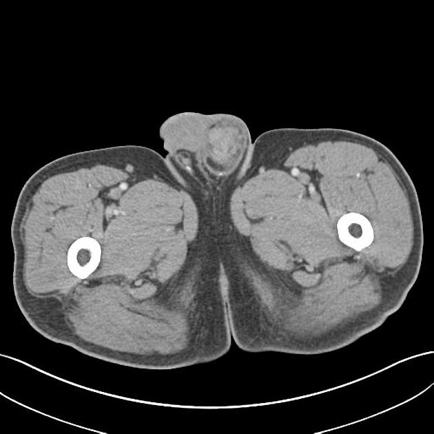
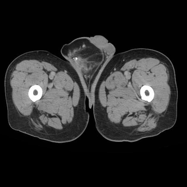
It is the small muscular sac that contains and protects the testicles. The spermatic cord is formed at the opening of the inguinal canal known as the deep inguinal ring. Fol lowing the path of the gonadal vessels confirms that the visualized structure in the ic represents either an ovary or a testicle. At ct testes are isoattenuating to soft tissue and show contrast enhancement.











:background_color(FFFFFF):format(jpeg)/images/article/en/vas-deferens/uVMWKLu6XA8k7X75k72Oug_lyembAetdHm1MrUfHZ6ZQ_Ductus_deferens_02-2.png)

